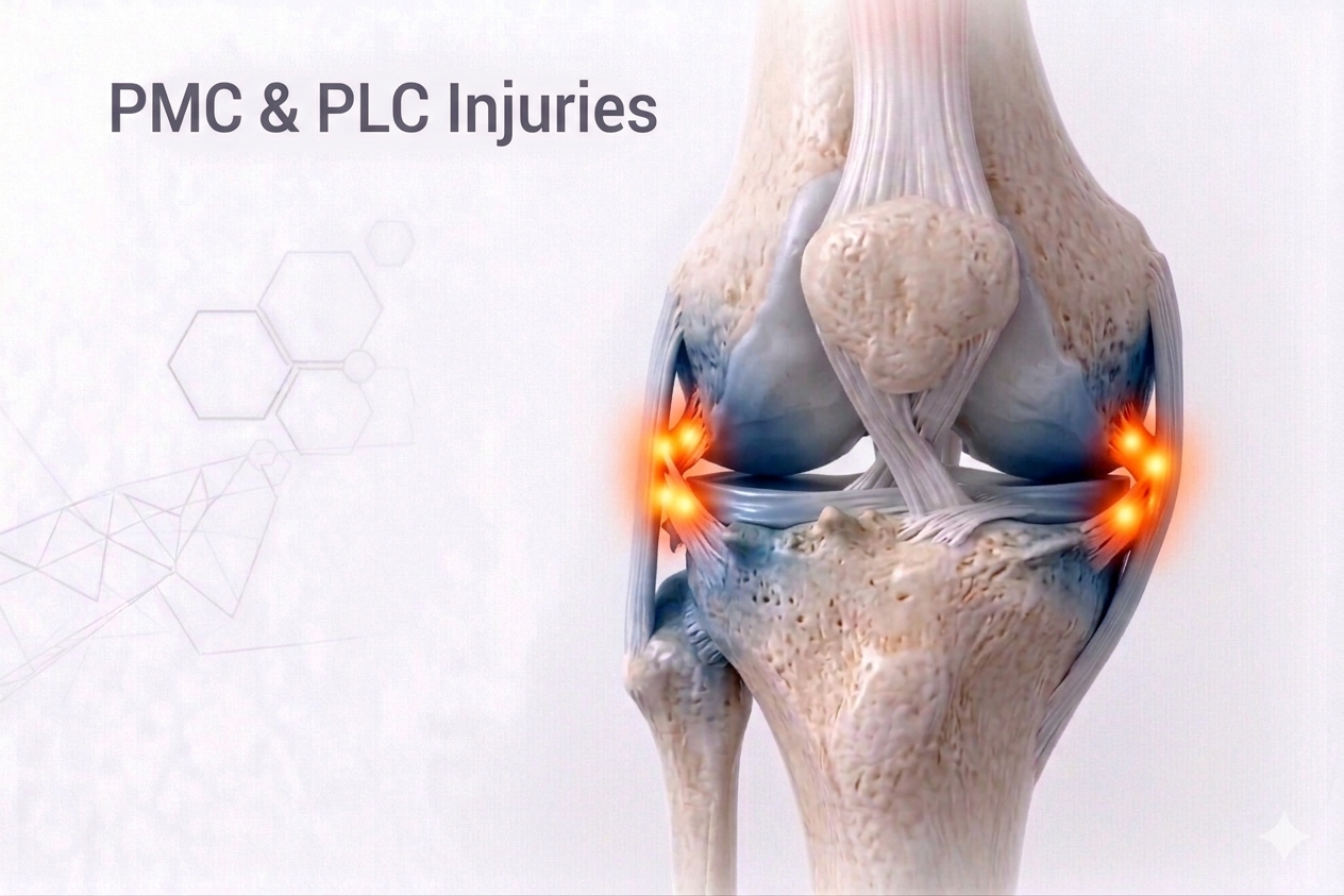
What Are PMC & PLC Injuries?
The Posteromedial Corner (PMC) and Posterolateral Corner (PLC) of the knee are complex anatomical regions composed of multiple ligaments, tendons, and capsule structures that work together to maintain rotational and translational stability.
- The PMCresists valgus and external rotation stress.
- The PLCresists varus and external rotation forces.
Injuries to these corners often occur in association with ACL or PCL tears, leading to multidirectional instability, difficulty walking downhill, or a feeling that the knee “twists out” during motion.
These are commonly missed injuries and require a high index of suspicion for timely diagnosis.
⚙️ How Do They Develop?
Posteromedial Corner (PMC) Injury
- Caused by a valgus forceor external rotation to a flexed knee.
- Often associated with MCL tearsor PCL injuries.
- Mechanism: Sudden inward collapse of the knee during contact or a fall.
Posterolateral Corner (PLC) Injury
- Caused by varus stress, hyperextension, or external tibial rotation.
- Common in road traffic accidents, contact sports, or falls with knee hyperextension.
- Often coexists with PCL or ACL tears(“knee dislocation spectrum”).
⚠️ Structures Involved
🩸 Posteromedial Corner:
- Superficial Medial Collateral Ligament (sMCL)
- Deep MCL
- Posterior Oblique Ligament (POL)
- Posteromedial Capsule
- Semimembranosus expansion
⚙️ Posterolateral Corner:
- Lateral Collateral Ligament (LCL)
- Popliteus Tendon and Popliteofibular Ligament
- Arcuate Ligament Complex
- Lateral Capsule
- Biceps Femoris Tendon
🔬 Pathophysiology
Both PMC and PLC act as secondary stabilizers to the cruciate ligaments (ACL & PCL).When injured:
- PMC injuriescause increased valgus and external rotation laxity.
- PLC injuriesresult in varus instability, hyperextension, and external tibial rotation (“dial test” positive).
If unrecognized, these injuries increase strain on ACL or PCL grafts, leading to recurrent failure after reconstruction— one of the most common causes of revision surgery.
🧪 Investigations
- Clinical Examination:
- PMC Injury:
- Tenderness on medial side.
- Increased valgus laxity in flexion.
- Positive “Anteromedial Drawer Test.”
- PLC Injury:
- Tenderness on lateral side.
- Dial Test Positive(increased external rotation at 30° & 90°).
- Posterolateral Drawer Testor “Reverse Pivot Shift” positive.
- May present with foot drop due to common peroneal nerve injury(in PLC).
- PMC Injury:
- Imaging:
- X-rays:Stress views to detect gapping or subluxation.
- MRI:Gold standard for identifying ligament tears, capsule injury, and concurrent ACL/PCL involvement.
- CT (3D):For avulsion or fracture evaluation.
💊 Management
🩹 Non-Surgical (For Mild, Isolated Sprains)
- Immobilization:Hinged brace or immobilizer for 3–4 weeks.
- Physiotherapy:
- Quadriceps activation and range-of-motion exercises.
- Avoid rotational and varus–valgus stress initially.
- Gradual strengthening:Emphasis on hamstrings and proprioceptive training.
🩺 Surgical (For Complete or Combined Injuries)
- Indications:
- Grade III injuries.
- Combined cruciate + corner injuries.
- Chronic instability after failed ligament reconstruction.
Surgical Options:
- Anatomic Reconstruction:Using tendon grafts (semitendinosus, biceps femoris, or allograft).
- Avulsion Fixation:For bony avulsions at fibular head or tibial attachment.
- Combined ACL/PCL + Corner Reconstruction:Performed simultaneously to restore multi-planar stability.
- Nerve Decompression or Neurolysis:For associated peroneal nerve palsy.
Rehabilitation:
- Protected weight-bearing with brace for 6 weeks.
- Early passive motion within safe range.
- Progressive strengthening from 8–10 weeks.
- Return to sport at 8–10 months with neuromuscular retraining.
⏳ Sequelae if Left Untreated
- Chronic rotational or varus/valgus instability
- ACL/PCL graft failure (if associated injury missed)
- Progressive medial or lateral compartment arthritis
- Recurrent knee dislocation or giving-way episodes
- Peroneal nerve palsy (in PLC)
- Poor functional outcomes and limited return to spor
🌟 Prognosis
With accurate diagnosis and anatomic reconstruction, outcomes are excellent — over 85–90% of patients regain full stability and functional performance.Failure rates drop dramatically when PLC or PMC injuries are recognized and treated alongside cruciate reconstructions.
💬 Key Takeaway
“PMC and PLC injuries are the hidden saboteurs of knee stability — often missed, but never harmless.Identifying and treating them alongside cruciate injuries ensures a strong, balanced, and lasting reconstruction.”