What is a PCL Injury?
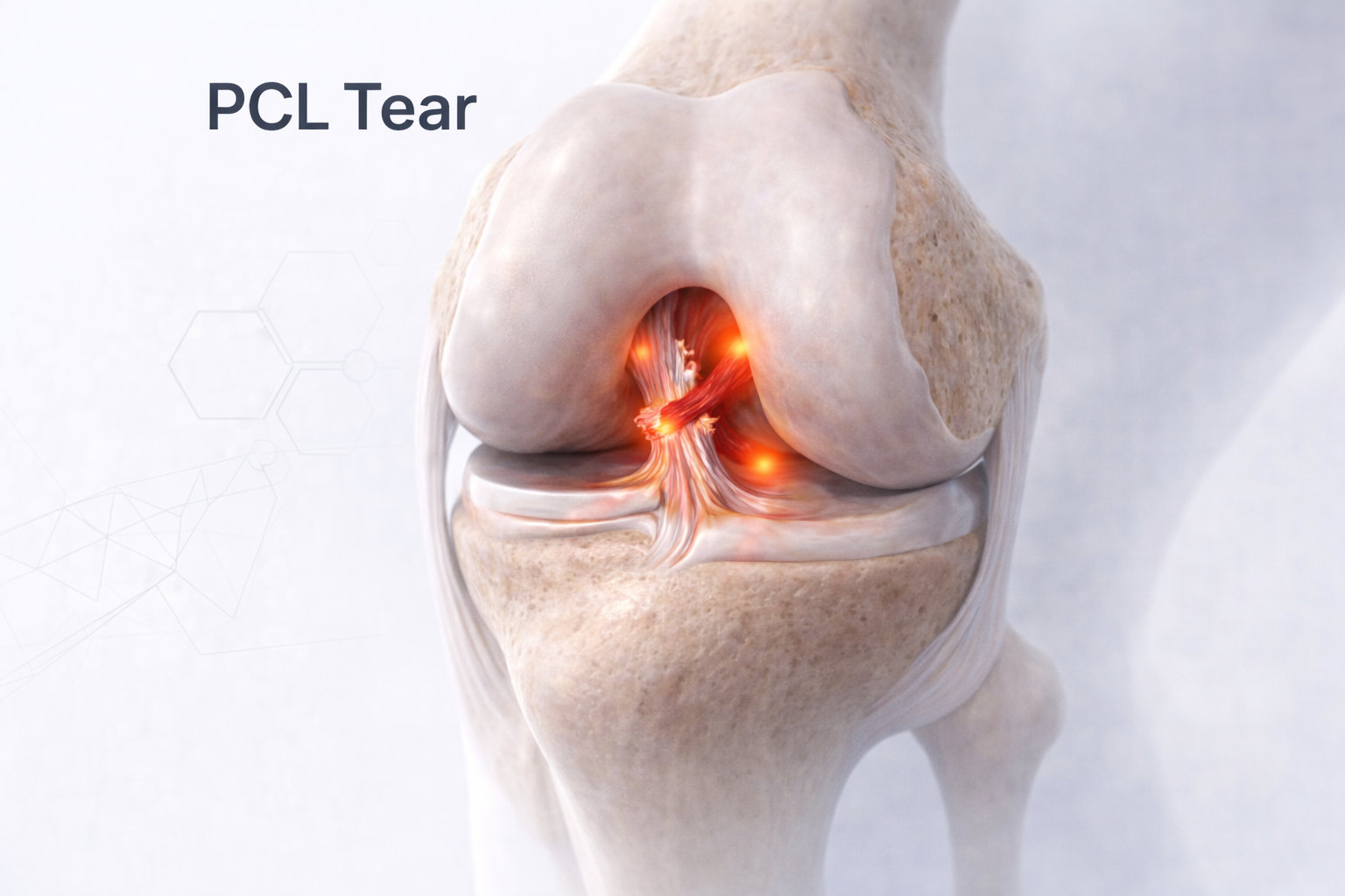
The Posterior Cruciate Ligament (PCL) is one of the key stabilizing ligaments of the knee, located just behind the ACL.
It prevents the tibia (shin bone) from moving too far backward relative to the femur (thigh bone) and plays a vital role in maintaining posterior and rotational stability.
A PCL Injury occurs when this ligament is stretched or torn — commonly due to a direct blow to the front of the tibiaor a fall on a bent knee.
Although less common than ACL injuries, PCL tears can cause significant pain, instability, and early joint wear if untreated.
⚙️ How Does it Develop?
The PCL can be injured in several ways:
- Direct Impact: Dashboard injury during a car accident — when the shin hits the dashboard with the knee bent.
- Sports Trauma: Fall or collision in sports like football, cricket, or rugby.
- Overextension: Landing awkwardly from a jump or sudden deceleration.
- Combined Ligament Injury: Often occurs with ACL or collateral ligament tears in high-energy trauma.
⚠️ Risk Factors
- Contact sports or road traffic accidents
- Falls on bent knees
- Weak hamstrings or poor lower limb coordination
- Previous knee ligament injury
- Improper rehabilitation post ACL or meniscus surgery
🔬 Pathophysiology
The PCL is thicker and stronger than the ACL and consists of two main bundles — anterolateral and posteromedial.
Injury leads to:
- Posterior translation of tibia, resulting in a “sagging” knee appearance.
- Altered knee biomechanics, increasing pressure on the medial and patellofemoral compartments.
- Secondary degenerative changes, particularly if unrecognized or untreated.
🧪 Investigations
- Clinical Examination:
- Posterior Sag Sign: Tibia appears sunken back when knee is bent at 90°.
- Posterior Drawer Test: Increased backward movement of the tibia.
- Pain, swelling, and feeling of instability or “looseness” while descending stairs.
- Imaging:
- X-ray: May show posterior tibial translation or bony avulsion.
- MRI: Gold standard for confirming PCL tear, associated meniscal or chondral injuries.
💊 Management
🩹 Non-Surgical (For Partial or Isolated Low-Grade Tears)
- Rest & Immobilization: Knee brace maintaining slight forward tibial translation.
- Physiotherapy:
- Quadriceps strengthening (to pull tibia forward).
- Avoid early hamstring strengthening (can worsen posterior sag).
- Focus on proprioception and stability training.
- Activity Modification: Avoid deep squats or heavy lifting during recovery.
🩺 Surgical (For Complete or Multiligament Tears)
- Arthroscopic PCL Reconstruction:
- Torn ligament replaced using a tendon graft (hamstring, quadriceps, or allograft).
- Graft fixed anatomically within femoral and tibial tunnels.
- PCL Repair (Selective): In acute avulsions with good tissue quality.
- Combined Reconstruction: In multiligament injuries involving ACL, MCL, or posterolateral corner.
- Rehabilitation:
- Brace with posterior support for 6–8 weeks.
- Early range-of-motion and quadriceps activation.
- Sports-specific training by 5–6 months.
- Full return to activity by 9–12 months.
⏳ Sequelae if Left Untreated
- Persistent instability (“giving way” feeling)
- Progressive cartilage wear and early osteoarthritis
- Medial or patellofemoral joint degeneration
- Difficulty descending stairs or running downhill
- Chronic pain and loss of confidence in the knee
🌟 Prognosis
With appropriate surgical reconstruction and dedicated rehabilitation, over 85–90% of patients regain full stability and function.
Isolated partial tears respond very well to structured physiotherapy.
Long-term outcomes are excellent when alignment and muscle balance are properly restored.
💬 Key Takeaway
“A PCL tear may not grab headlines like an ACL — but left untreated, it quietly destabilizes your knee.
Early diagnosis and balanced rehab ensure a strong, steady comeback.”