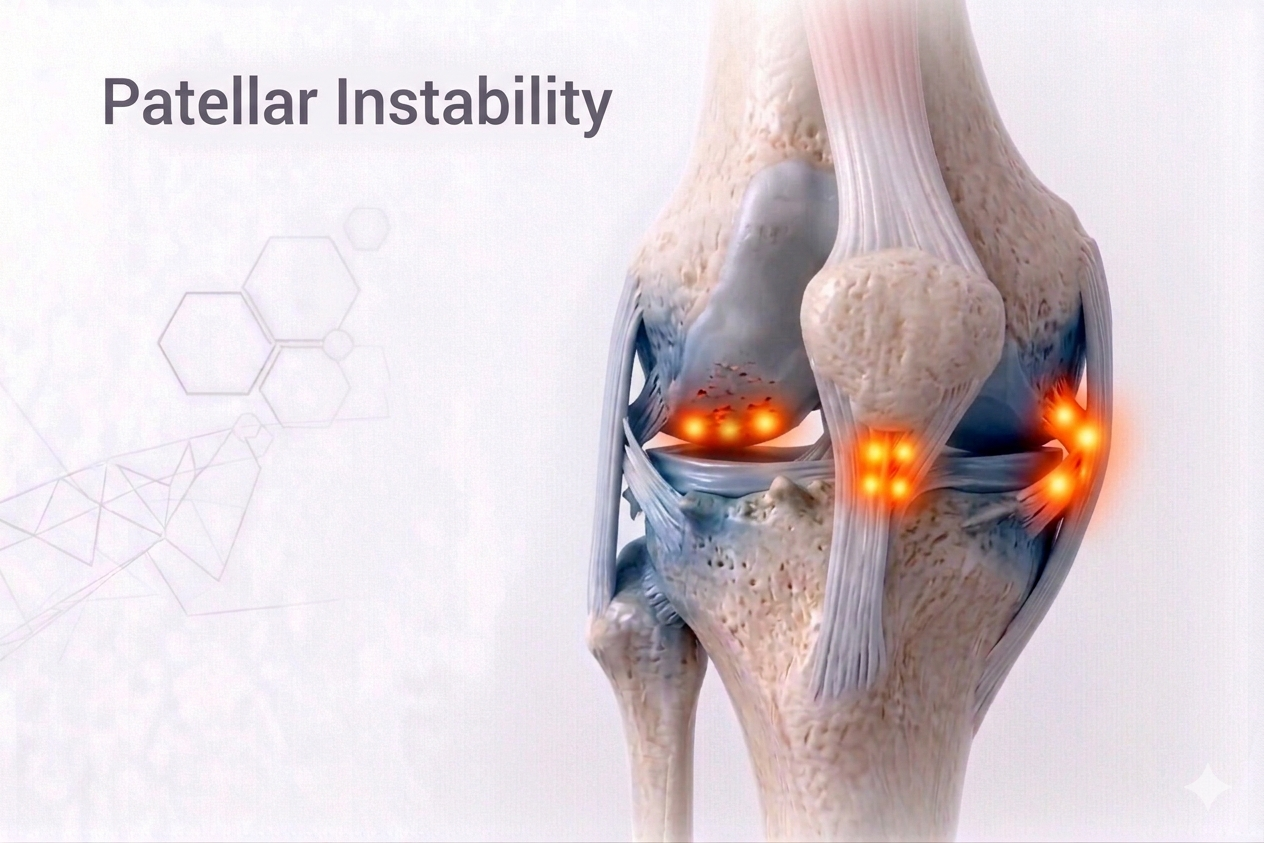
What is Patellar Instability?
The patella (kneecap) sits in a groove at the lower end of the thigh bone (femur), called the trochlear groove.During knee motion, it glides smoothly within this track — guided by muscles, ligaments, and bone alignment.
Patellar Instability occurs when this tracking is lost and the patella moves abnormally — partially (subluxation) or completely (dislocation) out of the groove.It causes pain, swelling, giving way, and fear of movement. Recurrent dislocations may lead to cartilage injury and early arthritis if untreated.
⚙️ How Does it Develop?
Patellar dislocation usually happens when the knee twists inward with the foot planted and the quadriceps contracts suddenly.The kneecap slips laterally (towards the outer side) — often accompanied by a “pop” and immediate pain or swelling.
In chronic cases, repeated episodes occur even with minor activities like running or stair climbing.
⚠️ Risk Factors
- Shallow trochlear groove (trochlear dysplasia)
- High-riding patella (patella alta)
- Increased Q-angle(knock knees, flat feet)
- Ligamentous laxity (loose ligaments, especially in females)
- Weak quadriceps (especially Vastus Medialis Obliquus – VMO)
- Family history or previous patellar dislocation
🔬 Pathophysiology
The Medial Patellofemoral Ligament (MPFL) and surrounding soft tissues stabilize the kneecap medially.In a traumatic dislocation:
- The MPFL ruptures,
- The patella moves laterally, and
- Cartilage on the patella or trochlea may get damaged.
Chronic instability results from failure to heal properly or underlying anatomical malalignment.
🧪 Investigations
- Clinical Examination:
- Positive Apprehension Test(patient feels knee will pop out when patella pushed laterally).
- Tenderness over medial patella border.
- Swelling or visible lateral shift after dislocation.
- Imaging:
- X-rays (AP, Lateral, and Skyline views):Show patellar tilt or high patella.
- MRI:Evaluates MPFL tear, cartilage injury, and trochlear morphology.
- CT Scan (optional):Measures TT–TG distance (tibial tubercle–trochlear groove offset) to assess malalignment.
💊 Management
🩹 Non-Surgical (First-Time or Mild Instability)
- Immobilization:Short-term brace or patellar stabilizing sleeve.
- Physiotherapy:
- Strengthening of VMO (Vastus Medialis)and core muscles.
- Balance and proprioceptive retraining.
- Stretching of tight lateral structures (IT band, lateral retinaculum).
- Taping or Bracing:To maintain patellar alignment during activity.
- Activity Modification:Avoid deep squats and sudden twisting until stability improves.
🩺 Surgical (For Recurrent or Anatomical Abnormalities)
- MPFL Reconstruction:
- Gold-standard procedure to restore medial soft-tissue restraint.
- Performed using hamstring or synthetic grafts.
- Tibial Tubercle Osteotomy (TTO):
- Repositions the patellar tendon attachment to correct tracking (used when TT–TG distance >20 mm).
- Trochleoplasty:
- Deepens the trochlear groove in severe dysplasia cases.
- Lateral Release:
- Loosens tight lateral structures (used selectively).
Rehabilitation:
- Protected movement for 4–6 weeks in a brace.
- Strengthening and dynamic control training for 3–4 months.
- Return to sport at 5–6 months depending on recovery.
⏳ Sequelae if Left Untreated
- Recurrent dislocation or subluxation
- Cartilage damage and early patellofemoral arthritis
- Weakness and instability during activity
- Chronic anterior knee pain
- Reduced performance and confidence
🌟 Prognosis
Modern MPFL reconstruction and alignment correction techniques offer excellent long-term outcomes, restoring confidence and function in >90% of patients.Early rehabilitation and attention to biomechanical alignment are essential for lasting success.
💬 Key Takeaway
“A slipping kneecap isn’t just a minor inconvenience — it’s a sign of imbalance.Precision diagnosis and stabilization restore not just your knee, but your confidence to move freely again.”