What are Collateral Ligament Injuries?
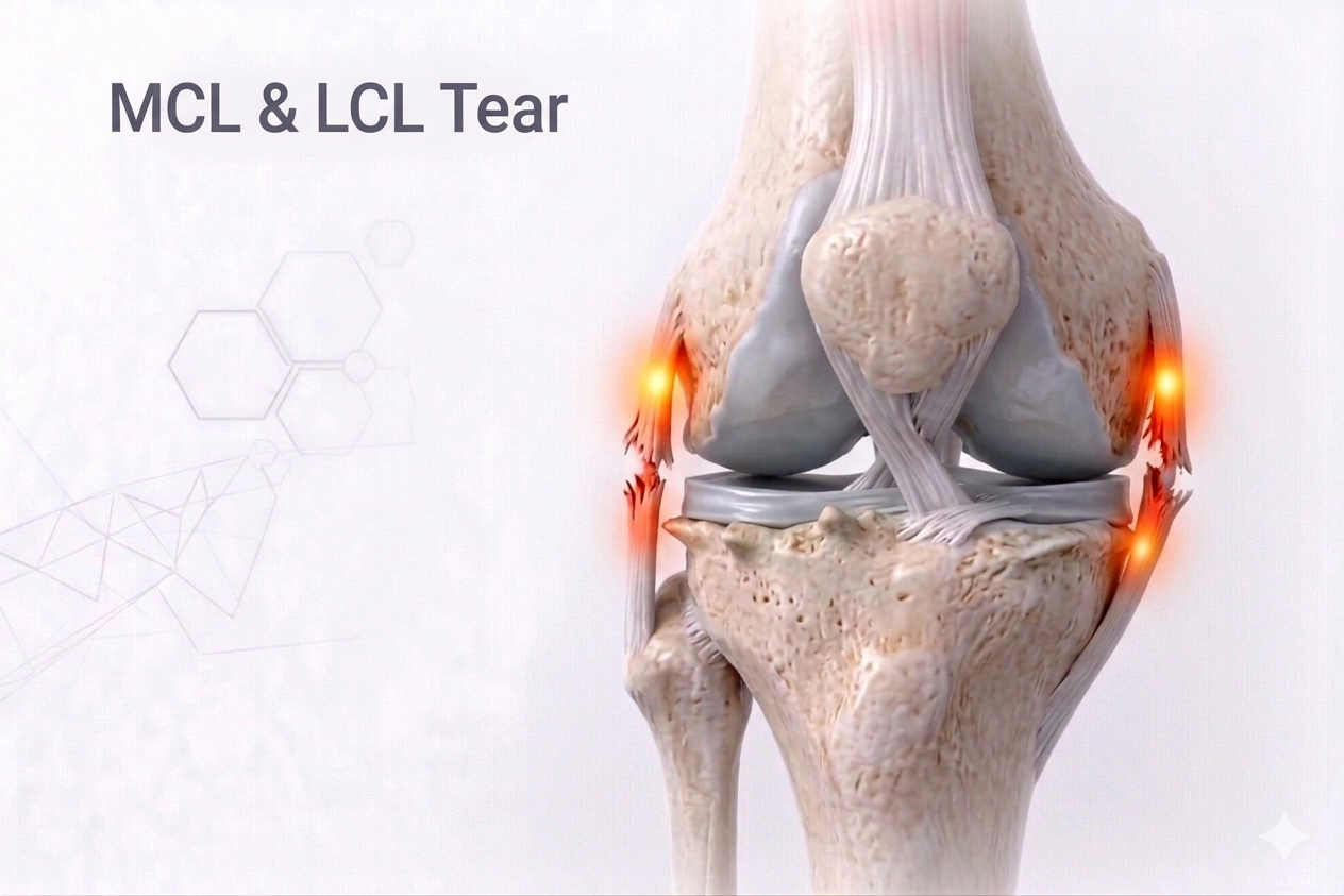
The collateral ligaments are strong bands of tissue on either side of the knee that provide side-to-side stability:
- MCL (Medial Collateral Ligament)— on the inner side, resists inward (valgus) stress.
- LCL (Lateral Collateral Ligament)— on the outer side, resists outward (varus) stress.
An MCL or LCL injury occurs when the knee is forcefully pushed sideways, stretched beyond its normal range, or twisted during activity.These injuries range from mild sprains to complete tears and are common in contact sports or after a fall or awkward landing.
⚙️ How Do They Develop?
- MCL Injury:Usually caused by a blow to the outside of the knee (valgus force), driving it inward — common in football or hockey.
- LCL Injury:Occurs from a blow to the inside of the knee (varus force), pushing it outward — often associated with high-energy trauma or multi-ligament injuries.
- Both may also occur with ACL or PCL tearsin combined ligament injuries.
⚠️ Risk Factors
- Contact or collision sports
- Sudden twisting while the foot is planted
- Poor landing mechanics during jumping
- Weak hip and core stability
- Previous knee injuries or ligament laxity
🔬 Pathophysiology
The MCL and LCL stabilize the knee during valgus and varus stress respectively.Injury leads to:
- Microscopic fiber disruption (Grade I)→ Pain and tenderness but no instability.
- Partial tear (Grade II)→ Increased laxity with a firm endpoint.
- Complete tear (Grade III)→ Significant instability, often involving adjacent structures like the meniscus or cruciate ligaments.
Prolonged instability can alter knee biomechanics and accelerate cartilage wear, predisposing to early arthritis.
🧪 Investigations
- Clinical Examination:
- MCL:Pain along the inner knee; tenderness over the medial joint line; positive valgus stress test.
- LCL:Pain on the outer knee; positive varus stress test.
- Swelling, bruising, and difficulty bending the knee.
- Imaging:
- X-ray:To rule out associated fractures or avulsions.
- MRI:Confirms ligament tear grade, extent, and associated ACL/PCL or meniscus injuries.
💊 Management
🩹 Non-Surgical (Most MCL & Mild LCL Injuries)
- Rest, Ice, Compression, Elevation (RICE)during the acute phase.
- Bracing:Hinged knee brace for stability during healing.
- Physiotherapy:
- Early range-of-motion exercises to prevent stiffness.
- Quadriceps and hamstring strengthening.
- Balance and proprioceptive training.
- Return to Sports:
- 3–4 weeks for Grade I,
- 6–8 weeks for Grade II,
- 10–12 weeks for Grade III (isolated).
🩺 Surgical (For Severe or Complex Tears)
- Indications:
- Complete (Grade III) tears with gross instability.
- Combined ligament injuries (ACL/PCL + MCL/LCL).
- Avulsion fractures of ligament attachment.
- Procedures:
- Primary Repair(for acute avulsions) or Reconstruction (using grafts).
- Posterolateral Corner Reconstructionfor severe LCL injuries.
- Rehabilitation:
- Gradual weight-bearing in a hinged brace.
- Full motion and strengthening by 10–12 weeks.
- Return to competitive sports in 4–6 months depending on associated repairs.
⏳ Sequelae if Left Untreated
- Persistent medial or lateral instability
- Early cartilage degeneration and osteoarthritis
- Chronic pain or swelling with activity
- Poor performance in pivoting or cutting movements
- Secondary ligament or meniscus damage
🌟 Prognosis
Isolated collateral ligament injuries heal very well with conservative care.Surgical reconstruction in complex cases also achieves >90% return-to-sport rates, provided structured rehab is followed.Modern bracing and early mobilization protocols prevent stiffness and recurrence.
💬 Key Takeaway
“Not every knee injury means surgery — MCL and LCL tears often heal beautifully with precision diagnosis, smart bracing, and guided rehab.”