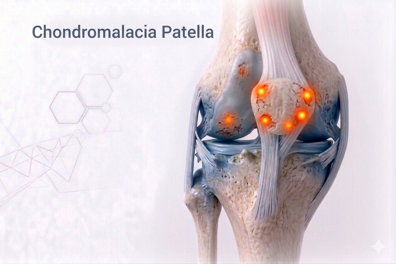
What is Chondromalacia Patella?
Chondromalacia Patella, commonly known as Runner’s Knee or Patellofemoral Pain Syndrome (PFPS), is a condition where the cartilage on the underside of the kneecap (patella) becomes softened, irritated, or damaged.It causes pain in the front of the knee, especially during activities like climbing stairs, running, squatting, or prolonged sitting.
Although often seen in athletes, it can affect anyone — particularly young adults and office workers who sit for long periods.
⚙️ How Does it Develop?
The kneecap glides within a groove on the femur called the trochlear groove.If the patella is misaligned or the muscle balance around the knee is disturbed, abnormal pressure develops on one part of the cartilage, leading to friction, inflammation, and softening.
Over time, this causes pain, roughness, and eventually cartilage wear — progressing from Chondromalacia to early Patellofemoral Arthritis if ignored.
⚠️ Risk Factors
- Malalignment of the patella (high-riding or lateral tilt)
- Weak quadriceps, especially Vastus Medialis Obliquus (VMO)
- Tight lateral structures (IT band or lateral retinaculum)
- Flat feet or abnormal gait mechanics
- Overuse (running, jumping, squatting)
- History of patellar instability or trauma
- Poor ergonomics or prolonged sitting (“movie-goer’s knee”)
🔬 Pathophysiology
The patellar cartilage undergoes repetitive microtrauma due to uneven tracking.This leads to:
- Softening (Grade I)
- Fissuring or fragmentation (Grade II–III)
- Exposed bone (Grade IV – advanced chondromalacia)
Persistent maltracking further irritates the synovium, causing inflammation and anterior knee pain.
🧪 Investigations
- Clinical Examination:
- Pain behind or around the kneecap.
- Worsens while climbing stairs, running, or sitting with bent knees.
- Crepitus (“grinding” sensation) on movement.
- Positive Patellar Compression Testor Clarke’s Sign.
- Imaging:
- X-ray (Sunrise / Skyline View):Evaluates patellar alignment.
- MRI:Detects cartilage softening, fissuring, and associated inflammation.
- Gait / Biomechanical Assessment:Useful in recurrent or sport-related cases.
💊 Management
🩹 Non-Surgical (Mainstay of Treatment)
- Activity Modification:Avoid deep squats, kneeling, or stairs during acute pain.
- Physiotherapy:
- Strengthening VMO, hip abductors, and core muscles.
- Stretching of IT band, quadriceps, and hamstrings.
- Patellar tracking and taping techniques to realign the kneecap.
- Footwear Correction:Orthotic insoles for flat feet or pronation.
- Medications:NSAIDs or short-term anti-inflammatory gels.
- Injections:
- PRPor Hyaluronic Acid (HA) injections to improve cartilage health.
- Viscosupplementationfor persistent chondral pain.
🩺 Surgical (For Advanced or Refractory Cases)
- Arthroscopic Debridement or Chondroplasty:
- Smoothing of damaged cartilage and removal of inflamed tissue.
- Lateral Release:
- To correct excessive lateral pull on the patella.
- Tibial Tubercle Osteotomy (TTO):
- Realignment procedure to improve tracking.
- Cartilage Restoration Procedures:
- Microfracture, OATS, or MACIfor focal cartilage loss.
Rehabilitation:
- Early motion and gradual strengthening.
- Return to sports or running typically within 3–4 months (non-surgical) or 6 months (post-surgical).
⏳ Sequelae if Left Untreated
- Persistent anterior knee pain
- Recurrent swelling and crepitus
- Patellofemoral cartilage degeneration
- Development of patellofemoral arthritis
- Decreased performance and activity tolerance
🌟 Prognosis
With early intervention and guided physiotherapy, over 90% of patients improve without surgery.Athletes can expect full return to performance with muscle retraining and biomechanical correction.
Modern arthroscopic and biologic treatments offer durable results for advanced cases.
💬 Key Takeaway
“Most front-of-the-knee pain isn’t from overuse — it’s from imbalance.Strengthen right, align smart, and your knees will thank you for life.”