What is Osteoarthritis of the Knee?
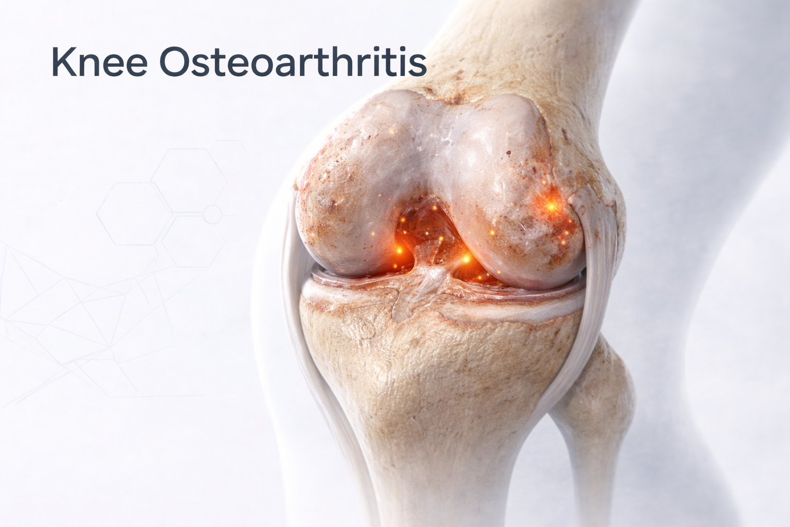
Osteoarthritis (OA) is a degenerative joint disease in which the smooth cartilage covering the ends of bones gradually wears away.
The knee is one of the most commonly affected joints, leading to pain, stiffness, swelling, and loss of mobility.
As the cartilage thins, the bones begin to rub directly against each other, forming bone spurs (osteophytes) and triggering inflammation.
The condition progresses slowly but can severely impact quality of life and independence if untreated.
⚙️ How Does it Develop?
Osteoarthritis develops due to imbalance between cartilage wear and repair.
Over time, factors like age, repetitive stress, previous injuries, or genetic predisposition accelerate cartilage breakdown.
As the protective layer deteriorates:
- The joint space narrows.
- The surrounding bone hardens and thickens.
- Inflammation of the joint lining (synovium) leads to pain and swelling.
⚠️ Risk Factors
- Age above 45 years
- Previous knee injuries (meniscus tear, ligament injury, fracture)
- Overweight or obesity (increased joint load)
- Repetitive squatting or occupational stress
- Family history of arthritis
- Malalignment (bow legs or knock knees)
- Post-meniscectomy or post-traumatic arthritis
🔬 Pathophysiology
In a healthy knee, cartilage acts as a cushion that absorbs impact and allows smooth motion.
In osteoarthritis:
- Cartilage degeneration exposes the subchondral bone.
- Synovial inflammation causes fluid accumulation (effusion).
- Muscle weakness and deformity (varus/valgus) develop.
This leads to a cycle of pain → disuse → stiffness → further degeneration.
🧪 Investigations
- Clinical Examination:
- Pain during walking, climbing stairs, or getting up from sitting.
- Morning stiffness (<30 minutes).
- Crepitus (grinding sound) during motion.
- Bony enlargement and limited range of motion.
- Imaging:
- X-rays: Joint space narrowing, osteophytes, subchondral sclerosis, deformity.
- MRI: Detects early cartilage damage or meniscal degeneration.
- Gait / Alignment Analysis: Determines load distribution and deformity pattern.
💊 Management
🩹 Non-Surgical (First-Line for Early to Moderate OA)
- Lifestyle Modification: Weight control and low-impact exercise (cycling, swimming).
- Physiotherapy:
- Strengthening of quadriceps and core.
- Range-of-motion and flexibility exercises.
- Bracing & Orthotics: Unloader braces for varus/valgus alignment.
- Medications:
- NSAIDs or analgesics for pain control.
- Glucosamine and chondroitin as supportive supplements.
- Injections:
- PRP (Platelet-Rich Plasma) or Stem Cell Therapy for cartilage regeneration.
- Hyaluronic Acid (Viscosupplementation) for lubrication.
- Corticosteroid Injections for short-term relief during flare-ups.
🩺 Surgical (For Advanced or Refractory Cases)
- Arthroscopic Debridement: For mild cartilage fraying or loose bodies (selected cases).
- High Tibial Osteotomy (HTO): For young, active patients with early medial compartment OA and varus alignment.
- Unicompartmental Knee Replacement (UKR): For single-compartment OA.
- Total Knee Replacement (TKR): Gold standard for advanced, multi-compartment arthritis.
- Restores alignment, stability, and pain-free motion.
Rehabilitation:
- Early walking post-surgery (within 24–48 hours).
- Strengthening and gait training from 2 weeks.
- Full recovery in 6–8 weeks with improved quality of life.
⏳ Sequelae if Left Untreated
- Persistent pain and stiffness
- Progressive deformity (bowing or knock-knees)
- Restricted mobility and difficulty walking
- Loss of muscle strength and balance
- Dependency on support for daily activities
🌟 Prognosis
With early diagnosis and multimodal treatment, pain relief and mobility improvement are achievable at all stages.
Modern surgical techniques and biologic therapies offer long-lasting outcomes with >95% patient satisfaction in total knee replacement.
💬 Key Takeaway
“Arthritis isn’t just age catching up — it’s a joint calling for attention.
With the right treatment, movement and independence can be beautifully restored.”